
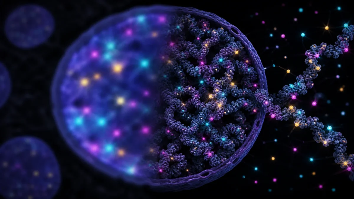
New fluorescent molecules can map chromatin at about 20-nanometre resolution in living cells and localize individual probes to roughly three nanometres in preserved cells—close to the two-nanometre width of the DNA double helix.
Published: August 30, 2026, 11:25 p.m. PKT · Reporting cutoff: August 30, 2026, 11:15 p.m. PKT
What you need to know
- The probes, called HoTs, enter cells, bind DNA and switch their fluorescence on and off without an added blinking buffer.
- STORM microscopy resolved chromatin to about 20 nanometres inside living cells.
- MINFLUX localized individual dyes to about three nanometres, but the sharpest result used chemically preserved cells.
- The probes worked in cultured cells and tissue sections, including samples from three people with bowel cancer.
- This is an imaging advance, not a validated cancer diagnostic or treatment.
Every human cell must pack roughly two metres of DNA into a nucleus only a few micrometres across. That packaging—chromatin—is not passive storage. Its changing three-dimensional structure helps control which genes a cell can use.
Researchers from the Centre for Genomic Regulation in Barcelona, City University of Hong Kong and Guangdong Provincial People’s Hospital designed a family of probes that makes this folding visible with unusual spatial and temporal detail. Their key trick is controlled blinking.
Why blinking produces a sharper image
If thousands of fluorescent labels glow at once, nearby signals overlap into a blur. A HoT probe alternates between bright and dark states. When only a sparse set of molecules is visible in each camera frame, software can estimate their positions and combine thousands of frames into one super-resolution reconstruction.
With stochastic optical reconstruction microscopy, or STORM, the team imaged chromatin inside living human skin cells and living HeLa cells at about 20-nanometre resolution—roughly ten times finer than the approximately 200-nanometre diffraction limit of conventional light microscopy.
Using MINFLUX on preserved laboratory cells, the researchers localized individual probe molecules to roughly three nanometres. That number describes localization precision, not necessarily the ability to resolve every structural feature exactly three nanometres apart. The distinction matters when comparing the result with DNA’s approximately two-nanometre diameter.
What the researchers saw in tissue
The probes also worked in tissue sections, including zebrafish retina and wax-preserved bowel samples from three cancer patients. In the human samples, chromatin appeared less compact in tumour cells than in nearby healthy tissue.
That observation is consistent with the broader idea that cancer can disrupt genome organization. But three patients are far too few to establish a diagnostic threshold, measure false positives or show whether chromatin structure predicts disease progression.
The team additionally trained an image-analysis system called AINU on living-cell images. It distinguished skin cells from stem cells with reported accuracy between 96% and 98%. Those cell-classification results do not demonstrate that the system can diagnose cancer in patients.
What the probes cannot yet do
HoTs bind across the genome. They reveal the shape, density and movement of chromatin, but they do not independently identify a particular gene or DNA sequence. Researchers can combine the broad chromatin signal with other targeted methods, yet that adds complexity.
The sharpest MINFLUX measurements also required preserved material. Living cells were imaged at lower resolution with STORM. Phototoxicity, dye behavior across different tissues and the repeatability of quantitative measurements will all matter if the method moves from research microscopes toward clinical pathology.
How other headlines framed the finding
- The Centre for Genomic Regulation used the “self-blinking fairy lights” analogy and emphasized resolution near the width of DNA.
- EurekAlert foregrounded both live-cell imaging and possible future cancer applications.
- Agencia SINC stressed the unusual DNA-imaging resolution while explicitly noting that the probes label the whole genome.
Why the advance matters
A probe that works in living cells and standard preserved tissue gives researchers a bridge between motion and fine structure. They can watch chromatin change, then examine related architecture in archived samples prepared by familiar pathology workflows.
Bottom line: HoT probes provide a powerful new view of DNA packaging, reaching about 20 nanometres in living cells and three-nanometre localization in preserved cells. Claims about cancer diagnosis remain future possibilities, not clinical results.
Sources
- Wang et al., HoT Auto-Blinking Probes, Molecular Cell, August 28, 2026.
- Centre for Genomic Regulation research explanation.
- PubMed record for the study.
Editorial disclosure: The lead image is a concept illustration inspired by super-resolution microscopy, not experimental data or a patient scan. SciQuest received no payment to cover this research. To report a possible error, contact SciQuest.