

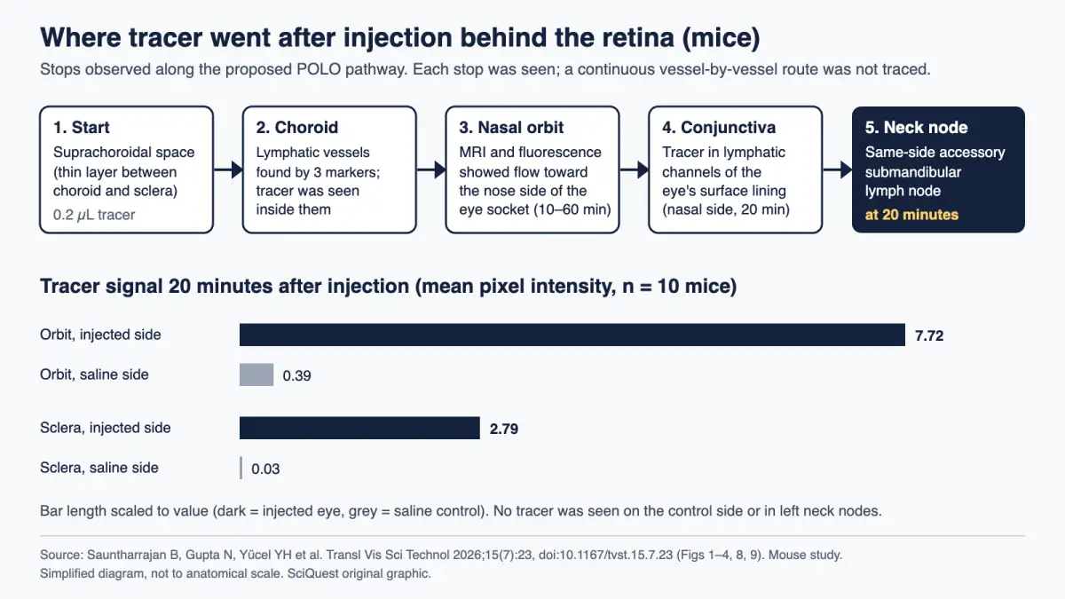
In 53 mice, tracer injected into a thin layer behind the retina reached a lymph node in the neck within 20 minutes, what the authors call the first direct evidence of a lymphatic exit from the back of the eye, though nobody has yet looked for it in people.
Mouse study · Peer reviewed
Researchers at the University of British Columbia and the University of Toronto have mapped a drainage route that carries fluid out of the back of the eye and into the lymphatic system, the body’s network for clearing excess fluid and waste. The team, led by ophthalmologists Neeru Gupta and Yeni Yücel, named it the posterior ocular lymphatic outflow (POLO) pathway and reported it in Translational Vision Science & Technology. It matters because glaucoma and macular degeneration are associated with fluid and waste build-up at the back of the eye. The catch: every result comes from mice.
What you need to know
- Finding: Mice have lymphatic vessels in the choroid, the blood-rich layer under the retina, and fluid placed behind it drains toward the nose side of the eye socket and on to a neck lymph node.
- Method: The team injected 0.2 microlitres of albumin-based tracer and followed it with 7-tesla MRI, near-infrared imaging and microscopy.
- Source: Sauntharrajan, Gupta, Yücel and colleagues, Translational Vision Science & Technology, 17 July 2026.
- Why it matters: It gives researchers a measurable exit route to study in eye diseases where fluid builds up, and a possible target for drug delivery.
- Limitation: It has not been shown in human eyes, and the authors did not trace an unbroken vessel route or test what happens when it is blocked.
Evidence at a glance
| Item | Assessment |
|---|---|
| Evidence status | Mouse study · Peer reviewed (received 1 September 2025, accepted 21 March 2026) |
| Primary source | Identification and Mapping of a Posterior Ocular Lymphatic Outflow (POLO) Pathway Through Choroidal Lymphatics, Transl Vis Sci Technol 15(7):23, published 17 July 2026, DOI 10.1167/tvst.15.7.23 (free full text) |
| Evidence strength | Preliminary to moderate for anatomy in mice; preliminary for disease relevance, because several independent methods agree on the mouse anatomy, but the work is from one lab group, has not been replicated, and did not study diseased eyes or people. |
| Principal limitation | Only mice were studied. Whether human eyes drain the same way, and whether this route changes in glaucoma, is unknown. |
| Coverage at a glance | 2 distinct publishers with their own write-ups found (SciTechDaily, Earth.com), as of 12 October 2026, 11:17 UTC. Counted: outlets that wrote their own article. Excluded: the universities’ own releases and verbatim reposts of the UBC release (ScienceDaily, News-Medical, Life Sciences BC). A quick search, not an exhaustive count. |
Source: the paper and the UBC/University of Toronto releases listed under Sources.
Is this a big deal?
It is a solid step in basic eye biology, not a medical advance. The eye has long been described as lacking the lymphatic drainage found in most organs, and the same two researchers began chipping at that idea in 2009 with a lymphatic route at the front of the eye. This study extends the idea to the back of the eye with direct experimental evidence. Whether it matters for patients depends on human studies that have not been done.
What scientists found
The researchers found vessels in the mouse choroid that carry three standard lymphatic markers at once: the proteins podoplanin and VEGFR-3, and the gene switch Prox1. In reporter mice that make Prox1-expressing cells glow red, the markers overlapped by 77% to 81%. By contrast, podoplanin overlapped by only 16.9% with a marker for ordinary blood vessels, which suggests these are a separate vessel type.
Tracer injected behind the retina turned up inside those choroidal lymphatic vessels, then in the tissues on the nose side of the eye socket, in lymphatic channels of the conjunctiva (the thin lining over the white of the eye), and finally in one lymph node under the jaw on the same side. The left side, which received saline, showed no tracer.
How they found it
The experiment was a tracking exercise. Under anaesthesia, the team made a tiny opening in the right eye and pumped 0.2 microlitres of tracer into the suprachoroidal space, the thin gap between the choroid and the white outer wall of the eye (the sclera). At 10 nanolitres per second, that injection took about 20 seconds.
The tracers were albumin, the most common protein in blood, tagged either with gadolinium for MRI or with a near-infrared dye. Albumin is a sensible stand-in for the protein-rich fluid that leaks from blood vessels, which lymphatics normally collect. Across all experiments the team used 53 mice from three strains, including albino mice whose lack of pigment let more light through for imaging.
MRI at 20 and 60 minutes (4 mice) and near-infrared imaging at 10 minutes (10 mice) both showed tracer moving toward the nose side of the orbit. At 20 minutes, fluorescence imaging of the neck found it in the right accessory submandibular lymph node. Under a hyperspectral microscope, tracer signal in the injected orbit averaged 7.72 units against 0.39 on the saline side, roughly 20 times higher (10 mice).
One check matters for interpreting the result. Pressure inside the eye dropped by about 9.6 mm Hg (about 1.3 kPa) after injection, but by the same amount in eyes given saline (9.56 vs 9.75 mm Hg, 8 mice). The authors attribute the drop to the anaesthetic, so the tracer itself did not appear to change eye pressure.
Why it matters to you
If you have glaucoma, or are at risk of it, this study changes nothing for you now. No test, eye drop or surgery is based on it, and it did not study glaucoma at all. Keep following your eye-care professional’s advice on pressure checks and treatment.
The longer-term interest is real. Glaucoma is closely tied to fluid balance inside the eye, and high eye pressure is a major risk factor for it. A newly mapped exit route gives researchers something to measure, and the UBC release says possible uses include better drug delivery to the back of the eye. Those are research directions, not results.
What the evidence does not show
The authors list their own limits plainly. MRI and fluorescence gave snapshots at set times, not continuous flow measurements. They did not show vessel-level continuity along the whole route, and they did not block any lymphatic segment to prove that drainage depends on it.
Three more gaps matter for readers. The study used healthy mice, so it says nothing about whether the route works differently in glaucoma or macular degeneration. Injected tracer is an artificial test, not the eye’s own fluid. Findings in mice do not always carry over to people, a point we also discussed in a mouse study on semaglutide and lifespan. And mouse eyes are far smaller than human eyes, so how much fluid this route handles in people, if it exists there, is unknown.
How this fits what we already knew
Doctors already know two exits for the clear fluid in the front of the eye (aqueous humour): a main drain near the edge of the iris and a slower “unconventional” route through surrounding tissue. Gupta and Yücel’s earlier work tied lymphatics to the front-of-eye route, reported that this drainage falls with age, and found that the glaucoma drug latanoprost increased it while timolol reduced it.
Lymphatic-like structures had also been reported in the choroid of monkeys and humans decades ago, the paper notes, but were not followed up. The new study is the first direct experimental evidence of a lymphatic route out of the back of the eye. That last claim is the authors’ own, and it awaits independent confirmation.
What other scientists say
We did not find published comment from independent researchers on this study. The quotations in coverage come from the study’s own authors. As Gupta put it in the UBC release, “This is a foundational discovery that shows the eye is not as closed a system as we previously thought.” Independent replication, ideally in human eye tissue, would be the real test.
How other headlines framed it
- ScienceDaily (a repost of the UBC release, 11 October) and SciTechDaily framed it as a newly discovered “cleanup system”, with treatment for blinding diseases as the payoff. Both mention the mouse-only evidence, but below the discovery angle.
- Earth.com led with the possibility of explaining vision loss, mentioning the mouse-only evidence briefly near the end.
- News-Medical ran the release as a waste-clearance finding with therapeutic promise, noting that human research is still needed.
None of the headlines said “mice”. Our headline does, because it is the single fact most likely to change how a reader judges the story.
What happens next
The authors propose time-resolved imaging such as photoacoustic tomography, tracers of different sizes, selective blocking of lymphatic segments and genetic mouse models. The UBC release says more work is needed to learn how the pathway works in humans. The open questions for glaucoma are whether the route exists in people and whether it changes with disease. We will cover results when they are published.
For a recent example of an eye therapy that did reach a person, see our report on doctors injecting mitochondria from a woman’s leg into her eyes, a single case with its own caveats.
In one sentence
In mice, fluid behind the retina has a lymphatic way out to the neck, which opens a new research question for glaucoma but gives patients nothing new yet.
Quick check: did it click?
- Where was the tracer injected? Into the suprachoroidal space, the thin gap between the choroid and the sclera at the back of the eye.
- How quickly did tracer reach a neck lymph node? Within 20 minutes, on the same side as the injected eye.
- Does this study change glaucoma treatment today? No. It was done only in healthy mice and did not test any treatment.
Read next
- Can Ozempic slow aging? What a mouse lifespan study can and cannot tell us
- How colour blindness works, and what the retina is doing
Sources and disclosure
- Sauntharrajan B, Gupta N, Zhou X, Gümeler E, Stanisz G, Lam WW, Koletar M, Yücel YH. Identification and Mapping of a Posterior Ocular Lymphatic Outflow (POLO) Pathway Through Choroidal Lymphatics. Translational Vision Science & Technology 2026;15(7):23. Published 17 July 2026. Open-access full text (PMC13390728).
- University of British Columbia Faculty of Medicine. Researchers discover the eye’s hidden cleanup system, 17 July 2026.
- University of Toronto Temerty Faculty of Medicine. Researchers discover eye’s hidden cleanup system, 21 July 2026.
- ScienceDaily. Repost of the UBC release, 11 October 2026.
Disclosure: This article was drafted with AI assistance and checked against the open-access paper and the university releases above. The 20-second injection time, the roughly 20-fold signal ratio and the kPa conversion (1 mm Hg = 133.3 Pa) are SciQuest calculations from the paper’s figures; the graphic is a SciQuest original drawn from the paper’s data. This article is not medical advice. Spotted an error? See our corrections policy.
Discover more from SciQuest
Subscribe to get the latest posts sent to your email.
Leave a comment