

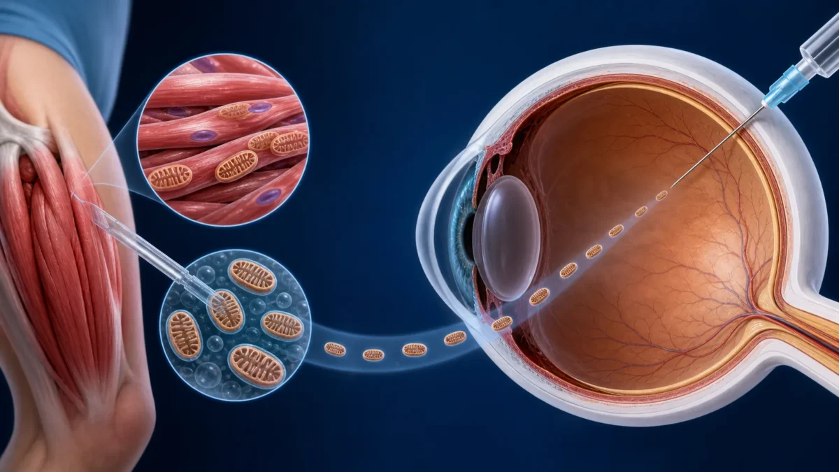
Doctors extracted functioning mitochondria from a 26-year-old woman’s thigh muscle and injected them into the vitreous fluid of her severely damaged eyes—the first documented procedure of its kind. The injections caused no detected ocular or systemic toxicity, and previously absent pupil responses briefly returned. Her measurable visual acuity did not improve. The report concerns one patient, has no control group and has not yet been peer reviewed.
Key facts
- The patient had severe bilateral optic neuropathy after prolonged loss of blood flow associated with a brain hemorrhage.
- Fresh mitochondria from her own skeletal muscle were injected into each eye 24 hours apart.
- No eye inflammation or systemic toxicity was detected in the case report.
- Pupil responses returned temporarily, but standard visual acuity did not measurably improve.
- The findings are a single-patient preprint, not evidence of an effective blindness treatment.
Why move mitochondria from muscle to the eye?
Mitochondria convert energy from food into adenosine triphosphate, the chemical energy cells can use. Retinal ganglion cells—the neurons that send visual information from the eye into the optic nerve—have unusually high energy demands. When blood flow and oxygen fall for too long, mitochondrial failure can contribute to cell injury and death.
Animal experiments have suggested that healthy mitochondria placed in the eye can be taken up by damaged retinal cells and may support their survival. The new procedure tested whether fresh, respiration-capable mitochondria could be delivered safely into a human eye.
Because the mitochondria came from the patient’s own tissue, the transplant was autologous: donor and recipient were the same person. That may reduce some immune risks, although injecting any biological material into the eye still carries serious concerns, including inflammation and infection.
What doctors did
The patient had lost vision in both eyes after prolonged cerebral hypoperfusion—insufficient blood flow to the brain—caused severe optic-nerve injury. Her condition had remained fixed for three months.
Under an emergency expanded-access authorization, clinicians took a muscle sample from her thigh and rapidly isolated fresh mitochondria at the point of care. They injected the preparation into the vitreous, the gel-like compartment behind the lens, first in one eye and then in the other 24 hours later.
The doctors monitored the patient for signs of intraocular inflammation, systemic reactions and changes in pupil behavior. Neither eye developed detected inflammation, and the report describes no ocular or systemic toxicity attributable to the procedure.
A biological signal returned, but vision did not
Before the transplant, automated measurements had recorded no pupil reaction across 45 readings over 71 days. Within days of each injection, pupil reactivity returned in the corresponding eye. The response was temporary and weakened after several weeks.
That signal is scientifically interesting because the pupil’s response to light depends on functioning retinal and neural pathways. It does not mean the patient regained useful sight. Standard testing found no measurable improvement in visual acuity. A later low-vision assessment suggested perception of shapes and shadows in one eye, but a single uncontrolled observation cannot show that the mitochondria caused it.
What this first case cannot tell us
A one-patient case report is designed to document feasibility and unexpected observations, not to establish efficacy. There was no untreated comparison eye, the two eyes were treated only a day apart, and recovery or measurement variation cannot be separated from a treatment effect.
The researchers also have not demonstrated that the injected mitochondria entered the patient’s retinal ganglion cells or remained functional there. The patient’s injury was severe and established, so many target cells may already have been beyond rescue. Different diseases or earlier treatment might produce different results, but that remains speculative.
The report is currently a preprint, meaning independent peer review has not been completed. Controlled studies would need to establish manufacturing standards, dose, delivery timing, durability, the patients most likely to benefit and both short- and long-term safety.
Bottom line: the procedure showed that a patient’s own mitochondria could be isolated and injected into the human eye without detected toxicity in this case. It did not restore measurable vision or prove that mitochondrial transplantation treats blindness.
Sources
- Putrino et al., “First intravitreal mitochondrial transplantation for bilateral vision loss,” Research Square preprint (2026).
- Miryam Naddaf, Nature news report and independent context, August 24, 2026.
- Nascimento-Dos-Santos et al., preclinical study of mitochondrial transplantation after optic-nerve injury.
Editorial disclosure: The lead image is a medical concept illustration, not a photograph of the procedure. SciQuest received no payment to cover this case. This article is informational and is not medical advice. To report a possible error, contact SciQuest.
Discover more from SciQuest
Subscribe to get the latest posts sent to your email.
Leave a comment